%%html
<script src="https://bits.csb.pitt.edu/preamble.js"></script>
<style>:root {--jp-cell-prompt-width: 0px}
.citation { font-size: small }
.reveal blockquote {
box-sizing: border-box;
width: 100%;
margin: 1em 0;
padding: 0.65em 1em;
font-size: 0.95em;
font-style: normal;
line-height: 1.4;
text-align: left;
color: #24344d;
background: #f0f4fa;
border-left: 5px solid #003594;
border-radius: 0 6px 6px 0;
box-shadow: none;
}
.reveal blockquote > :first-child {
margin-top: 0;
}
.reveal blockquote > :last-child {
margin-bottom: 0;
}
.pd-boltz p {
margin: 0;
}
.pd-pair {
display: grid;
grid-template-columns: 1fr 1fr;
gap: 22px;
align-items: center;
text-align: center;
}
.pd-pair img {
max-height: 405px;
width: auto;
}
.pd-figure {
text-align: center;
}
.pd-figure img {
max-height: 470px;
width: auto;
}
.pd-side .pd-visual {
min-width: 0;
text-align: center;
}
.pd-visual p {
margin: 0;
}
.pd-visual img {
margin: 0;
}
.pd-side .pd-visual img {
max-height: 480px;
width: auto;
}
.pd-side .pd-visual p {
margin: 8px 0;
}
.pd-side .pd-visual.pd-multiple img {
max-height: 220px;
}
</style>
Protein design: from an objective to a sequence
- Why design? Applications and useful properties.
- ProteinMPNN: choose a sequence for a backbone.
- RFdiffusion: generate a backbone with structural constraints.
Proteins are already medicines
Antibodies, enzymes and signaling proteins can themselves be therapeutics.

Annual CDER approvals. BLAs: biologics license applications; NMEs: new molecular entities.
Excludes products approved by CBER, including vaccines and gene/cell therapies.
How can we design their sequences and structures for the function we want?
Biocatalysis: a new scaffold around an active site

Crystal structure and design: win (rainbow/gray), PDB 9DEF. Fig. 3D.

Turnover: WT = parent win; dashed line = enzyme concentration. Shading: SD. Fig. 2E.
Design: RFdiffusion generates backbones; LigandMPNN designs sequences; Rosetta FastRelax refines models; PLACER assesses catalytic geometry.
Product exceeds enzyme concentration, demonstrating repeated turnover.
Vaccines: controlling antigen presentation

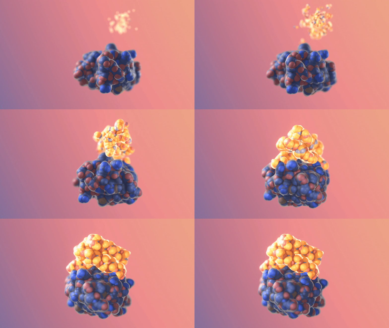
Structural model: blue RSV antigen on the gray/orange I53-50 scaffold at full valency. Fig. 1B.

Mouse serum neutralization: symbols = animals; bars = geometric means. Percentages = antigen valency. Fig. 4B.
Design: Rosetta-designed interfaces assemble I53-50; stabilized RSV F antigen (DS-Cav1) is displayed on its exterior.
Greater antigen display density gives higher neutralization titers in mice.
Antivirals: designed miniprotein inhibitors

Cryo-EM and design: LCB1 (magenta/gray) bound to RBD (cyan). Fig. 4B.

Neutralization: LCB1 and LCB3 reduce cultured-cell infection. Fig. 5B (2020 study).
Design: Rosetta docks miniprotein scaffolds and optimizes the RBD interface, followed by experimental screening and affinity optimization.
The small designed binder blocks a viral interaction needed for infection.
Antibodies: proteins that recognize specific targets

Immunoglobulin G (IgG) is a Y-shaped antibody composed of two heavy chains and two light chains.
- Fab (fragment antigen-binding): the two arms that recognize antigens.
- Fc (fragment crystallizable): the stem that interacts with immune receptors.
- Variable regions: determine which antigen the antibody recognizes.
- CDRs (complementarity-determining regions): six loops per binding site that largely determine binding specificity.
Nanobodies: single-domain antigen binders
Camelids produce heavy-chain-only antibodies. Their isolated variable domain (VHH, or nanobody) can recognize antigens without a light chain.
- Small (~15 kDa), typically stable and soluble.
- Three CDR loops form the antigen-binding surface.
- An elongated CDR3 can reach into pockets inaccessible to conventional antibodies.
Camelids
What do we design in an antibody?
- Epitope: the surface on the target (antigen) that we want to recognize.
- Paratope: the antibody surface that contacts it, usually involving the CDR loops.
- Design variables: CDR geometry and sequence, supported by a suitable framework.
- Molecular format: a VHH, a linked VH–VL fragment (scFv), or a full antibody
A useful binder also needs specificity, suitable expression and stability, and the biological effect required by its application.
Nanobodies: choosing the binding epitope

Cryo-EM VHH: navy; design: gray. VHH_flu_01 bound to HA (yellow), with glycans in teal. Fig. 3f.

SPR binding: blue = measured response; dashed black = global 1:1 fit. Fig. 2b.
Design: antibody-specialized RFdiffusion generates CDR backbones; ProteinMPNN designs their sequences; tuned RoseTTAFold2 filters complexes.
After experimental screening, VHH_flu_01 binds influenza HA with Kd = 78 nM.
Protein design beyond biomedical applications


Protein design
- Create proteins with specified properties.
- Redesign existing proteins to improve activity, stability, or specificity.
- Design can operate on sequence, structure, or both.

Protein sequence design with Rosetta
- Given a backbone, choose amino acids and side-chain conformations.
- Rosetta searches using a score function with physical and statistical terms.
- Packing, sequence search, and backbone relaxation can be combined.

Kuhlman · Designing protein structures and complexes with Rosetta · JBC (2019)
ProteinMPNN
- MPNN: message-passing neural network.
- Learns sequence preferences from protein structures.
- Supports fixed residues, multiple chains, and tied sequence positions.
- Generates sequences rapidly for downstream screening (faster than classical Rosetta).

The graph-based predecessor

- Ingraham et al. represent the backbone as a graph of spatial neighbors.
- An autoregressive decoder ("structured transformer") predicts residues from N to C in this earlier model.
- Graph attention/message passing makes nearby 3D context available during prediction.
$$p(\mathbf{s}\mid\mathbf{X})=\prod_i p(s_i\mid s_{<i},\mathbf{X})$$


Neighbors in space, not just sequence
- Residues far apart in sequence can be close in the folded structure.
- The structure encoder communicates over a spatial-neighbor graph.

ProteinMPNN: encode, then decode

- Backbone encoder: update residue and pair features.
- Sequence decoder: sample residues conditioned on the backbone and already assigned identities.
Autoregressive
For a decoding order $\pi$:
$$p(\mathbf{s}\mid\mathbf{X})=\prod_t p(s_{\pi_t}\mid\mathbf{s}_{\pi_{<t}},\mathbf{X}).$$
What changed in ProteinMPNN?
(Compared to previous MPNN approach)
- Use distances between N, Cα, C, O, and a virtual Cβ to describe local geometry.
- Update edge features as well as node features in the encoder.
- Use random decoding orders rather than always N to C.
- Evaluate the individual changes and their combination in ablations.
Decoding order and backbone noise
Random decoding order
- Allows known sequence positions to provide context for unknown positions.
- Supports multichain design and tied positions without a privileged N-to-C order.
Backbone noise during training
- Perturb coordinates to improve robustness to structural uncertainty.
Ablations
Accuracy: fraction of native amino acids predicted correctly.
Perplexity: $\exp(\text{mean per-residue cross-entropy})$; lower is better.
Bold score indicates training with Gaussian noise added to backbone coordinates

(Seeming) Aside: Single sequence structure prediction
Question: Why did AlphaFold2 and the original RoseTTAFold lose accuracy when predicting natural proteins from a single sequence?

Redesigned sequences are often easier to predict
- ProteinMPNN designed new sequences for 396 native backbones.

Rescuing hallucinated designs experimentally
- Structure-prediction-guided hallucination can produce sequences that express poorly.
- Redesigning the sequences for those backbones improved soluble expression in this test.
- Circular dichroism and temperature measurements test folding and thermal behavior.

Does the experimental structure match?
An X-ray structure of a redesigned protein closely matches its target backbone (2.35Å RMSD).

Designing sequences for nanoparticle assemblies
- Preserve the intended backbone and assembly geometry.
- ProteinMPNN generated 76 sequence designs for 27 tetrahedral backbones.
- 13 formed assemblies with the expected size in the reported experiments.

Rescuing Rosetta
- Scaffold a polyproline motif recognized by a Grb2 SH3 domain.
- ProteinMPNN rescued binding for designs that had failed with the original sequences.
- Disruptive mutations test whether the intended motif contributes to binding.

ProteinMPNN: what it contributes
- Rapid sequence proposals for monomers, assemblies, and motif-containing proteins.
- Learns sequence compatibility with a backbone from structural data.
- Reported example: 1.2s vs 258.8s for a 100-residue design on one CPU, compared with classic Rosetta protocol.

RFdiffusion overview
- Generate new protein backbone structures by denoising.
- Condition generation on motifs, target surfaces, or symmetry.
- Use ProteinMPNN to design sequences for those backbones.

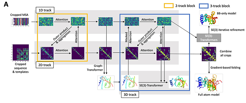
Starting from RoseTTAFold
- RoseTTAFold couples three representations:
- 1D: sequence/MSA features.
- 2D: residue-pair features.
- 3D: residue coordinates.
- RFdiffusion adapts the structure-prediction network to denoise protein structures.
Diffusion on protein structures
Training: corrupt known structures and learn to recover their clean geometry.
Generation: repeatedly denoise an initially random structure.

How the frames are noised and denoised
- Residues are represented by a frame:
- A translation $\mathbf{x}_i$: the Cα position.
- A rotation $R_i$: orientation of the N–Cα–C frame.
- Positions: Gaussian diffusion in 3D.
- Orientations: rotational diffusion on SO(3).
- The network predicts the clean structure from a noisy structure and its noise level.
- Train with unaligned mean-squared error on the reconstructed backbone coordinates.

Sampling a new protein backbone
- Initialize random residue positions and orientations.
- Predict a cleaner structure and take a reverse-diffusion step.
- Repeat while maintaining any specified constraints.


One generator, several design tasks
- Unconditional: create a plausible monomer.
- Symmetry: build a defined oligomeric assembly.
- Motif scaffolding: preserve a functional geometry.
- Binder design: complement a target surface.
ProteinMPNN then supplies sequences.
Self-consistency of generated monomers

Generate a backbone, design its sequence, then predict its structure with AlphaFold2.
Small design–prediction RMSD supports self-consistency.

Are the structures different from known ones?
- TM-score measures structural similarity; larger values indicate closer matches.
- Compare each design with its closest match in the PDB.

Symmetry as a generation constraint
- Specify a point-group symmetry and number of subunits.
- Jointly construct compatible subunits and interfaces.
- Applications include antigen-display particles and other designed assemblies.


Do the designed oligomers assemble?
Compare the design and predicted structure with negative-stain EM class averages and 3D reconstructions.

Motif scaffolding preserves functional geometry
- A motif is a set of residues with a desired spatial arrangement.
- Hold the motif fixed and generate a protein around it.
- Examples include catalytic residues or a binding epitope.

Testing motif-scaffolding methods
Success depends on preserving the motif and generating a compatible overall fold.


Designing a binder to a target surface

- Condition on the target structure and selected interface hotspots.
- Generate a complementary backbone, then its sequence.


A designed interface tested by cryo-EM
The HA_20–influenza hemagglutinin complex closely matches the intended binding geometry.
Structural agreement complements the binding assay.

RFdiffusion Summary
Generates candidate backbones for structural design tasks: unconditional, symmetry, motifs, binders.
Example: generating a binder for the insulin receptor.


LigandMPNN: protein design with atomic context
ProteinMPNN designs sequences from a protein backbone, but ignores nonprotein atoms.
LigandMPNN conditions sequence design on the protein backbone and surrounding atomic environment.

BoltzGen: a broader binder-design workflow
Jointly proposes sequence and all-atom structure, then refines and filters candidates.

Chroma: programmable generative protein design
- Integrated design: backbone diffusion + sequence and side-chain generation

Connecting sequence, structure, and function
- Sequence design: choose identities for a structure.
- Structure generation: choose geometry for a task.
- Co-design: propose sequence and structure together.

Key points
- Protein design starts with a desired function and definable constraints.
- Antibodies and nanobodies are binding scaffolds with different formats and engineering tradeoffs.
- ProteinMPNN proposes sequences conditioned on a backbone.
- RFdiffusion proposes backbones, including designs conditioned on motifs, targets, or symmetry.
- Structural confidence and self-consistency guide selection.
Journal Club
- Select paper to present today
- Groups of one or two
Assignment 7
- Due Wednesday, November 30
- Extra time provided to enable flexiblity, not procrastination
Project
- Proposals due Wednesday, November 11
- Groups of one to three (but higher expectations for groups of three)
- Poster presentation Wednesday, December 9
Exam 2
- Wednesday, December 2